At Brandeis University in Waltham, investments from the Massachusetts Life Sciences Center (MLSC) are expanding access to advanced imaging technologies that allow researchers to examine biological systems with greater precision and detail. By providing researchers with the tools to better understand how cells and tissues function, the MLSC’s support is helping advance discoveries that inform the development of new treatments and ultimately improve patient outcomes.
Through the MLSC’s Research Infrastructure program, Brandeis University’s Light Microscopy Facility has expanded its imaging capabilities with LifeCanvas Technologies’ SmartSPIM light sheet microscope, the Andor Dragonfly 620SR spinning disk microscope, and the ZEISS LSM 980 with Airyscan 2. Home to more than 13 pieces of high-power imaging equipment, the facility provides researchers with access to a broad range of specialized technologies, including these three state-of-the-art microscopes funded by the MLSC. Each of these microscopes provide researchers with powerful tools to study biological structures and processes, while making specialized imaging technologies more accessible to academic and industry investigators across the Commonwealth.
The LifeCanvas SmartSPIM microscope enables detailed three-dimensional imaging of biological tissues, allowing researchers to examine the organization and relationships of cells within larger samples. By providing a more comprehensive view of complex biological structures, the technology can help investigators better understand how tissues function and how they may be affected by disease.
Mike Vivian of the Grienberger Lab is investigating how distinct populations of neurons in the hippocampus contribute to learning and memory. By combining in vivo calcium imaging during behavior with light sheet imaging of cleared brains using the SmartSPIM, Vivian and his team can map neuronal activity alongside detailed three-dimensional neuronal structures. When integrated with molecular labeling, this approach connects neuronal function, structure, and gene expression at the single-neuron level, providing new insights into how cellular identity shapes memory circuits.
Complementing the SmartSPIM’s three-dimensional imaging capabilities, the Andor Dragonfly 620SR combines high-speed imaging with advanced resolution capabilities, allowing researchers to observe cellular activity and examine tissue samples that are often too thick for conventional spinning disk microscopes. The microscope also supports single-molecule localization microscopy at resolutions below 10 nanometers, along with advanced capabilities for optogenetics, laser ablation, and targeted stimulation, allowing researchers to examine cellular functions, interactions, and responses to changes in their environment.
Eratos Therapeutics, a startup company utilizing incubator space at Brandeis, is focused on advancing drug discovery for neurological and central nervous system disorders. The company is developing an AI-driven approach to nanoscale spatial mapping to identify potential therapeutic targets within native tissue, with applications in Alzheimer’s disease and other complex neurological disorders. By leveraging expansion microscopy, Eratos uses the Dragonfly 620SR to rapidly capture detailed images of expanded tissue at nanoscale resolution across large fields of view. These multichannel datasets are then integrated into the company’s AI pipeline to generate three-dimensional maps of protein targets, supporting the discovery of potential new treatments.
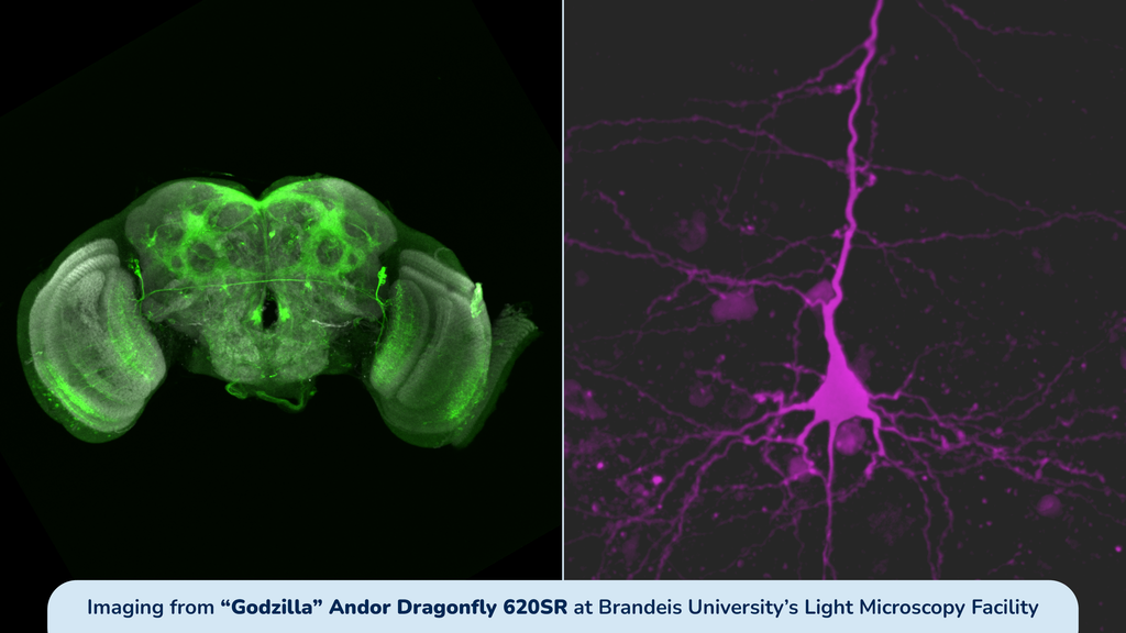
Further expanding the facility’s capabilities, the ZEISS LSM 980 with Airyscan 2 enables researchers to investigate biological systems at multiple scales, from the movement of individual molecules to the three-dimensional organization of tissues. Its advanced imaging technology allows researchers to distinguish overlapping fluorescent signals and capture fine cellular details at resolutions below 100 nanometers. The microscope also provides tools to measure molecular concentration, movement, and flow, helping investigators understand not only where molecules are located within cells, but also how they behave and interact. By connecting molecular activity with cellular structure, the technology offers new opportunities to study complex biological processes and advance discoveries related to health and disease.
Diane Bissen, PhD, and Masha Glyavina of the Turrigiano Lab are studying how learning changes neuronal circuits to optimize behavior. Using the LSM 980, they measure the size of dendritic spines in pyramidal neurons within the mouse visual cortex. These small, specialized protrusions on neuronal dendrites receive excitatory synaptic inputs and play an important role in communication between neurons. Because spine size closely correlates with synaptic strength, measuring changes in spine morphology provides a reliable indicator of synaptic plasticity. The LSM 980’s high-resolution imaging capabilities enable the researchers to precisely measure individual spines, offering valuable insights into how learning reshapes connections within the brain.
All three microscopes are accessible through the MLSC’s Research Equipment Database (R.E.D.), which connects companies, research institutions, and investigators with MLSC-funded equipment located at partner organizations across Massachusetts.
Applications for the MLSC’s Research Infrastructure program are open until this Friday, Nov. 6, 2026, at 1 p.m. EST. Click here to learn more and apply today.